- Frontotemporal Dementia affects ~15–22 per 100,000 people and is the most common early-onset dementia after AD — yet it takes on average 3+ years to correctly diagnose due to behavioral symptoms mimicking psychiatric disorders.
- Diffusion MRI provides white matter microstructure biomarkers that distinguish FTD from healthy controls and other dementias, but standard protocols demand 40+ gradient directions → prolonged scans impractical for cognitively impaired, agitated patients.
- Linear least squares (LLS) tensor fitting degrades severely below ~20 directions; a learning-based approach is needed to unlock clinically feasible sparse-dMRI protocols for FTD screening.
- A model achieving diagnostic accuracy from 5–21 directions could make dMRI-based FTD screening accessible in resource-limited settings and for patients who cannot tolerate full-protocol scans.
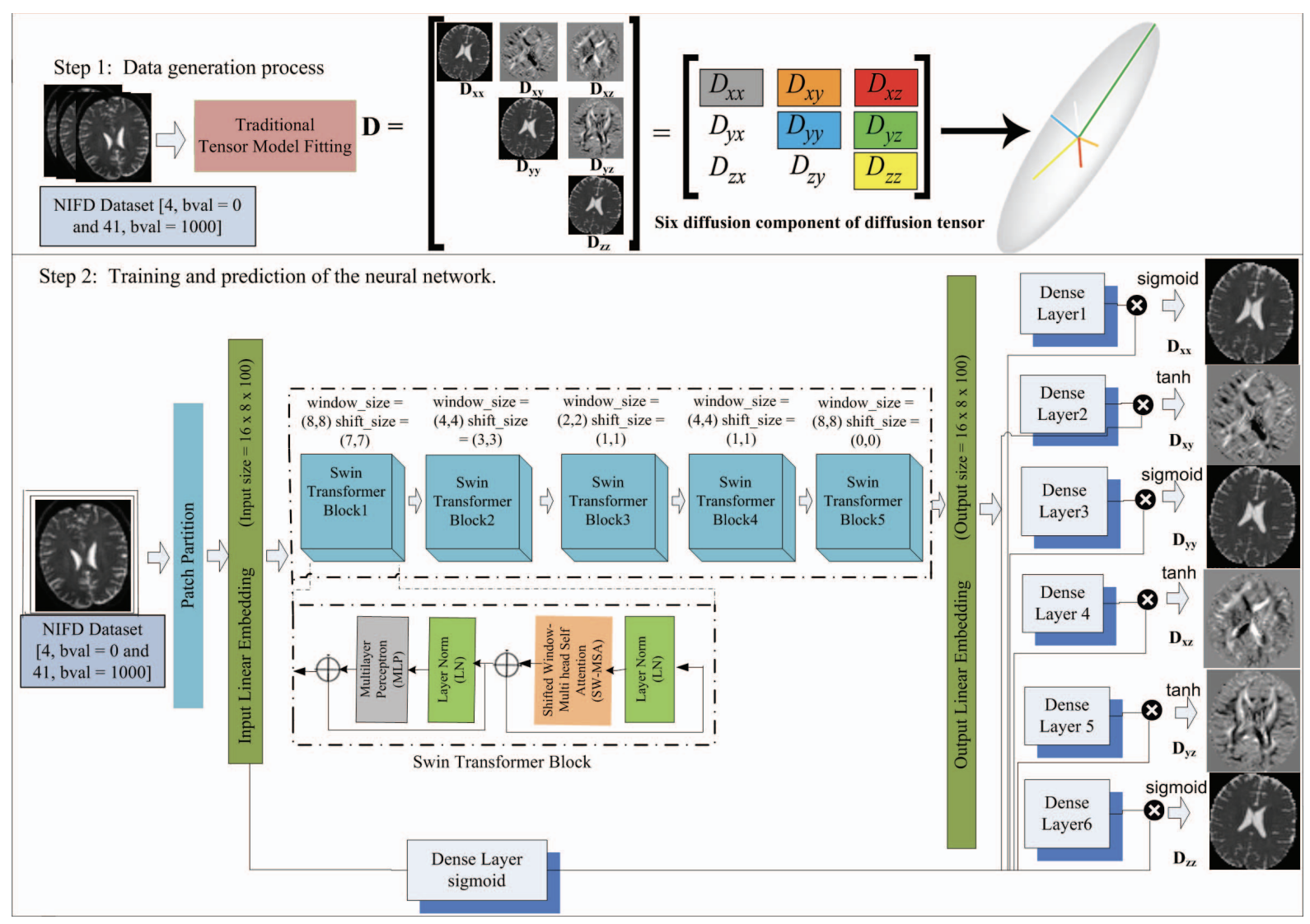
Pipeline: b=0 + sparse b=1000 DWI (5 or 21 directions) → Patch Partition (16×8 input) → 5 Swin Transformer Blocks with varied window/shift sizes → 6 parallel Dense Layers predicting diffusion tensor components (Dxx, Dxy, Dyy, Dxz, Dyz, Dzz) → FA, AxD, MD maps → TBSS analysis on NIFD white matter skeleton → FTD vs. healthy classification.
- First demonstration that sparse dMRI (5–21 gradient directions) is sufficient for reliable FTD diagnosis — reducing scan time by 50%+ while preserving diagnostic white matter biomarkers validated on the NIFD dataset.
- Swin-Transformer adapted for sparse DTI parameter estimation: shifted-window multi-head self-attention captures local voxel correlations across patch boundaries, enabling a single model to generalise across differing direction counts.
- Physics-informed loss function enforcing positive-definiteness and biophysical coherence in estimated diffusion tensors — preventing implausible anisotropy values at extreme sparse regimes (5 directions).
- Voxel-wise TBSS analysis on NIFD mapping FTD-specific white matter degradation in the uncinate fasciculus and superior longitudinal fasciculus — two tracts known to degenerate early in FTD — providing interpretable tract-level biomarkers from sparse acquisitions.
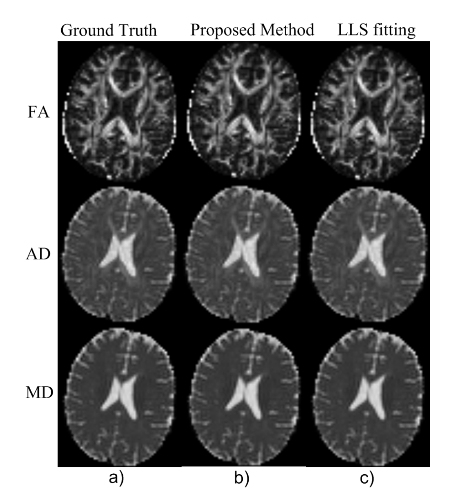
41 directions (full protocol): Proposed method matches ground truth FA, AxD, and MD maps and is comparable to full LLS fitting — validating reconstruction quality before testing sparse regimes.
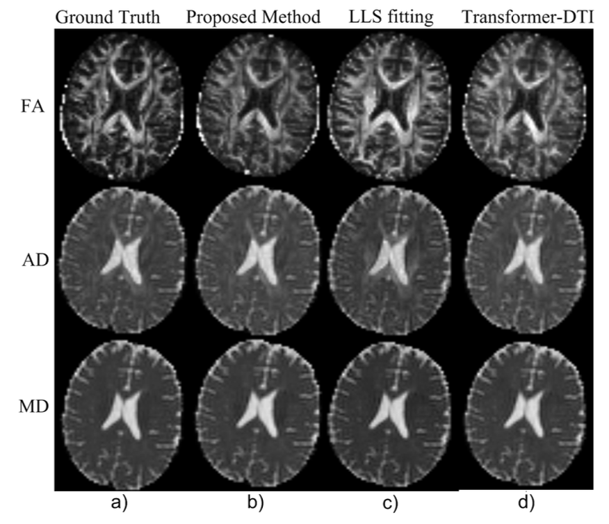
21 directions: Proposed method outperforms LLS and Transformer-DTI. FA structure well-preserved at half the standard directions.
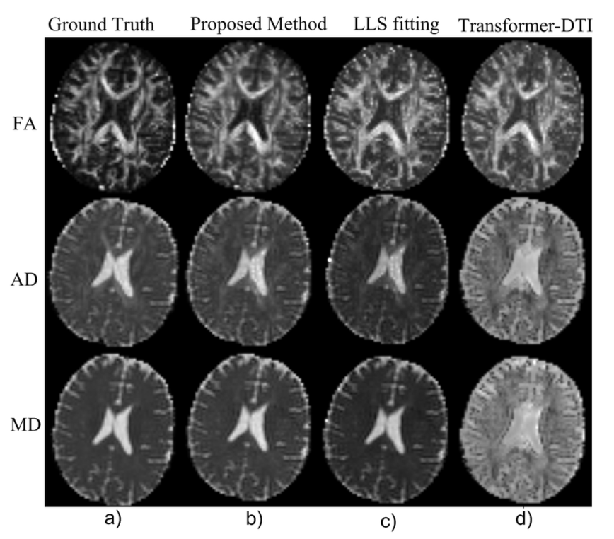
5 directions: Proposed surpasses both baselines. LLS produces severe artefacts; our model recovers clean diffusion maps.
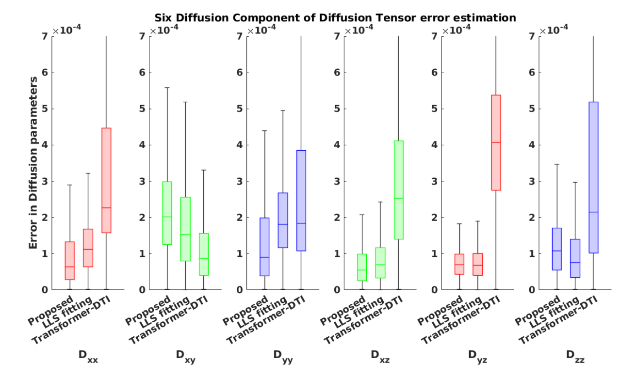
5 directions: Per-component error distributions. Proposed shows tightest spread across all 6 tensor components; LLS error variance is markedly higher.
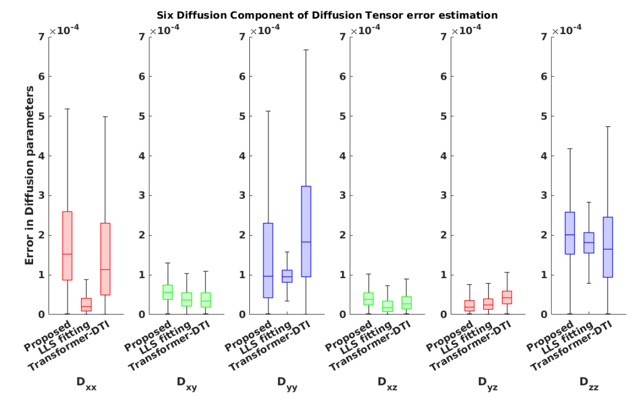
21 directions: All methods converge closer to ground truth, but Proposed retains a measurable accuracy advantage over both LLS and Transformer-DTI.
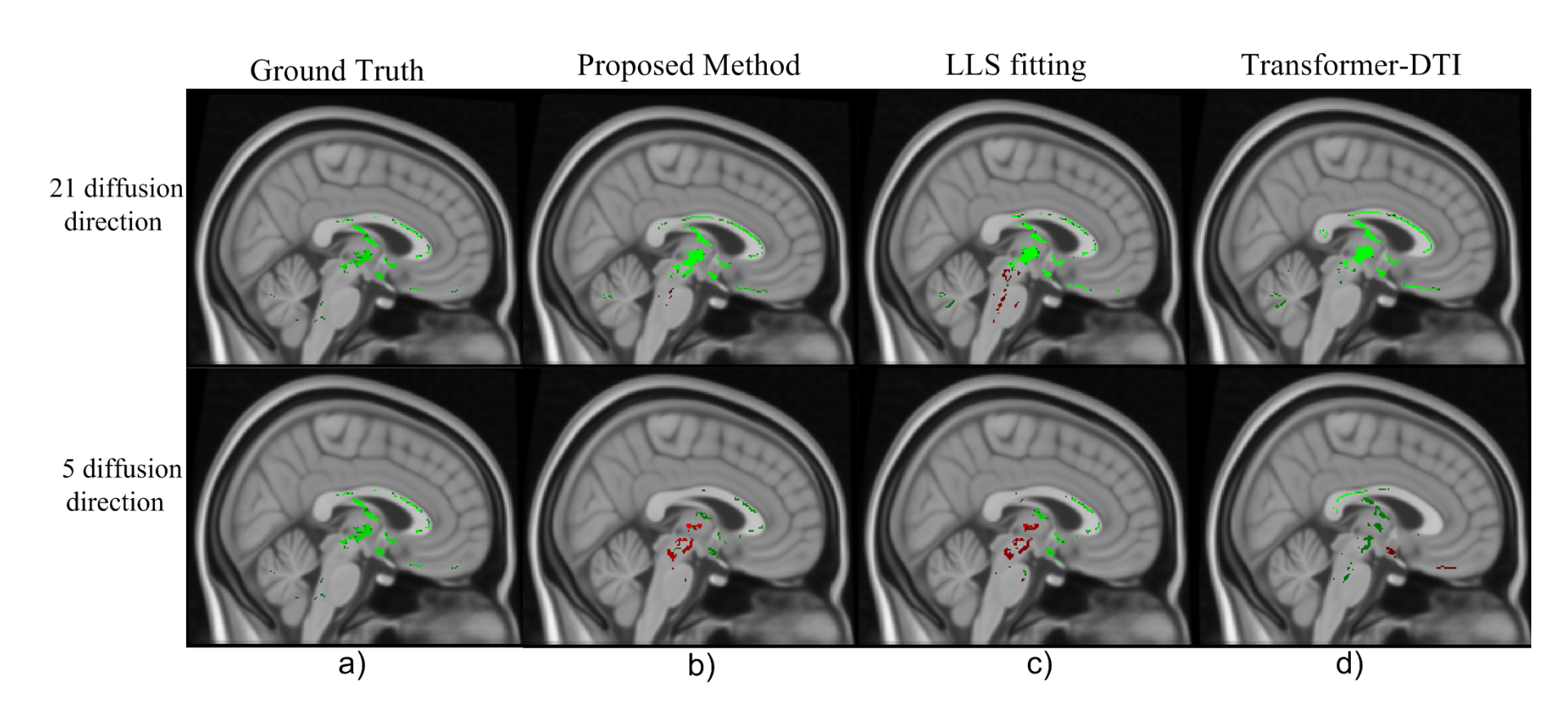
Cingulum (axial): Rows show 21-direction and 5-direction reconstructions. FA reductions in the cingulum bundle are preserved from sparse data — a key FTD biomarker tract.
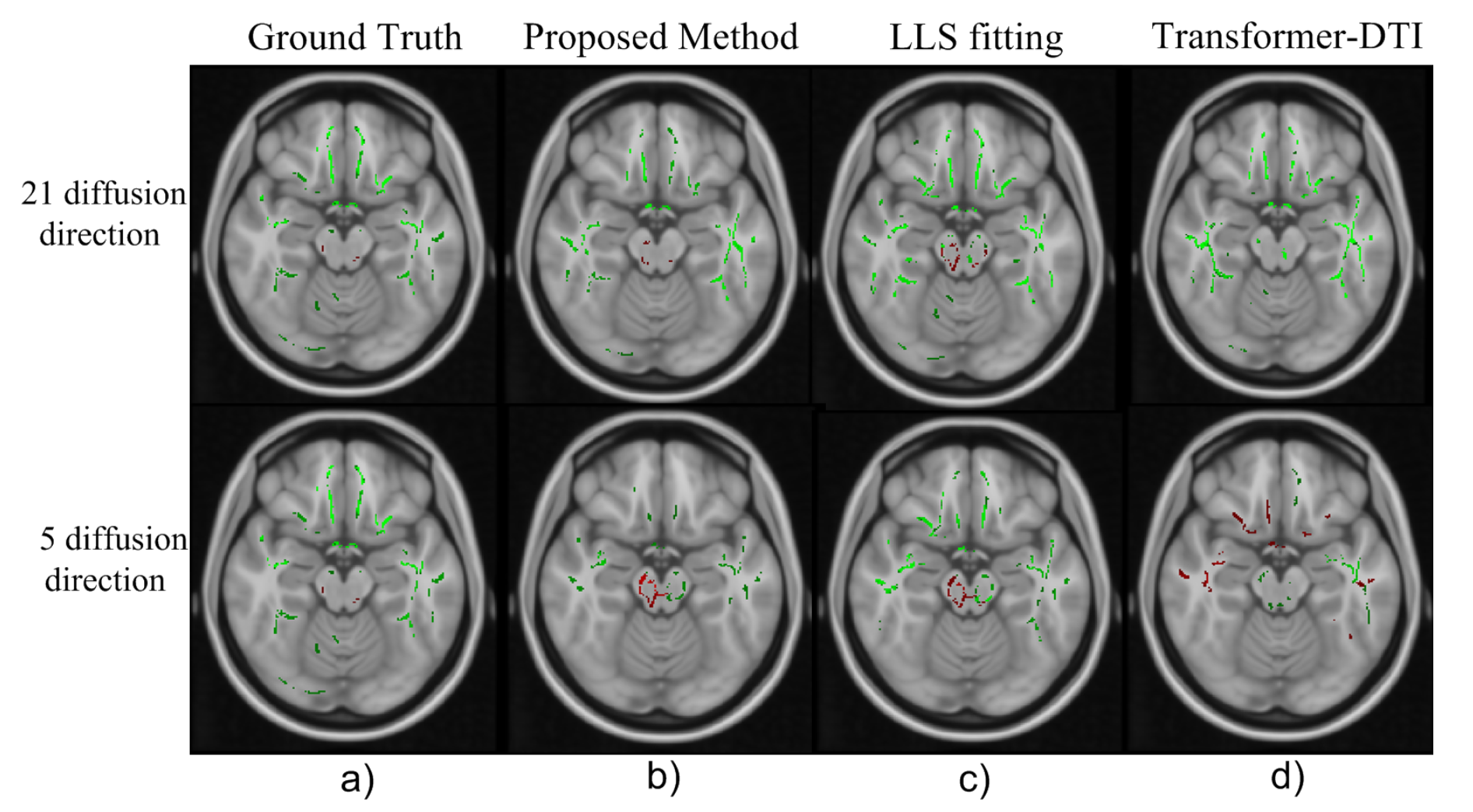
Uncinate Fasciculus (coronal): Temporal lobe white matter degradation linked to frontal-temporal disconnection in FTD. Proposed recovers the spatial signature from sparse acquisitions.